
Introduction to Arrhythmias - An Electrical Enigma
Our heart beats about 80 times every minute. That means that it beats about 100,000 times a day, everyday until we die. That seems a little insane does it not? But what is crazier is the fact that normally, every beat is exactly the same duration. 100,000 times a day. How does our heart maintain this precision? And does it ever go wrong?
Now in order for there to be a functional heart beat during which blood is effectively pumped out of the heart, the chambers need to pump in a sequence - the cardiac cycle. The cardiac cycle is the cycle of events that occur during every heart beat. It is exactly 0.8 seconds long and it consists of four events.
atrial contraction
atrial relaxation
ventricular contraction
ventricular relaxation

If you think about the heart in terms of electrical circuits, the two atria function in a parallel circuit and the two ventricles function in a parallel circuit but the atria and ventricles are connected in a series circuit with a slight delay in between. And the functioning of the heart, is indeed a complicated electrical circuit.

Our heart as an electrical circuit. Atria contract in parallel. Ventricles contract in parallel. But atria and ventricles contract in series.
The beginning of the cardiac cycle starts with the initiation of an electrical impulse. This impulse needs to conducted throughout the body of the heart, to every last myocyte and depolarise it. But the propagation of this impulse cannot be haphazard, it needs to be in perfect sequence in order to get the heart chambers to contract in the right order. And lastly, it needs to ensure that a second impulse does not start until the current impulse depolarises the whole myocardium.
This entire process is made possible by the conduction system of the heart.
The conduction system consists of the following parts organised according to rate of pacing of each tissue:
The Sino - Atrial Node (SA Node) -
The Atrio - Ventricular Node (AV Node) -
The bundle of His -
Purkinje Fibers -
Myocardium -

Electrical conduction system of the heart.
Reproduced from Grant’s Atlas Of Anatomy 13th Edition.
So the impulse is generated at the SA node, high up in the right atrium, because it is the fastest pacemaker. From here it is dispersed to the atria. While the atria are being depolarised, specialised pathways (preferential pathways) carry the impulse to the AV node as well. Here there is a pause. This pause allows the atria to contract fully and empty the blood into the ventricles. The impulses are then carried down the bundle of His and through the Purkinje fibers to the interventricular septum followed by the ventricular myocardium itself. This results in ventricular depolarisation.
There is a small concept that must be clarified before we talk further about the electrical activity of the heart. It is common knowledge that cardiac tissue is conductive tissue and that the myocytes themselves have the ability to propagate impulses. Why then, is there a need for a conduction system? Well nature’s beauty is apparent in the design of the heart. The atria and the ventricles are separated from each other by a set of fibrous rings which are electrically inert. This means that the only way for the electrical impulse to get from the SA node to the bundle of His is through the AV node. And the AV node is where the pause occurs. This is natures way of effectively allowing blood to go from the atria to the ventricle before the ventricles contract and send the blood off into the body.

Fibrous rings separating the atria from the ventricles.
Reproduced from Grant’s Atlas Of Anatomy 13th Edition.
Now these electrical impulses are documented on an electrocardiogram. This electrocardiogram, allows us to visualise the electrical impulses and also is our primary diagnostic tool to pin point any aberrancy in the cardiac rhythm. So let us see the normal waves on the ECG and what parts of the cardiac cycle they correspond to.

Normal Electrocardiogram.
Reproduced from Goldberger’s Clinical Electrocardiography 9th Edition
Now this is a normal ECG. There are a lot of things marked on this ECG, but right now, only five things are important.
P wave - denotes atrial depolarisation
PR interval - this is the pause that we spoke about. During this time, the heart is on hold, waiting for the blood to go from the atria to the ventricle.
QRS complex - denotes ventricular depolarisation. During this time atrial repolarization also occurs but is swallowed by the large ventricular depolarisation.
T wave - ventricular repolarization
RR interval - This denotes the total time taken for an entire cardiac cycle. It is a useful measurement to identify some arrhythmia.
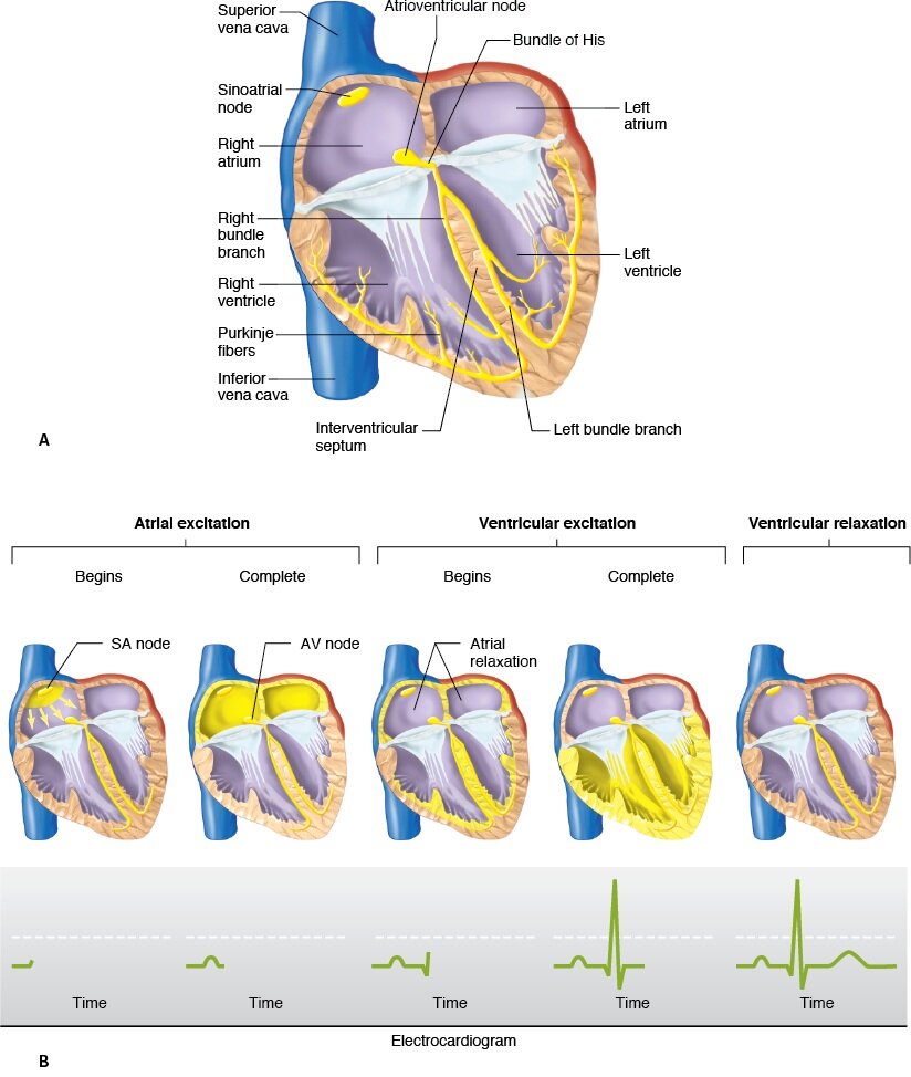
Illustration showing the cardiac conduction apparatus and the reflection of the electrical activity of the heart on ECG.
Reproduced from Ganong’s Review of Medical Physiology.
This is all you need to know about the normal electrical functioning of the heart in order to understand and identify arrhythmia.
Now arrhythmias are a confusing and complicated subject. They are a source of confusion to even some seasoned cardiologists. But understanding the basic concepts of arrhythmias will allow you to understand the intricacies of the pathologies.
Arrhythmias are basically a change in the rate or the rhythm of the beating heart.
They are classified based on where they originate.
Sinus arrhythmias
Supraventricular tachycardias
Ventricular tachycardias
We are going to discuss these separately.
Author: Narendran Sairam (Facebook)
Sources and citations