
Hypertrophic Cardiomyopathy - When too much is bad for you
You’ve all probably heard of news where a prominent, young athlete dies suddenly and unexpectedly while playing on the field. Breaking news suggests they died from sudden cardiac arrest. Autopsy reports show that the ventricles of the heart are massively hypertrophied and the ventricular cavity barely made out. Studies have found that anywhere from 8% to 40% of sudden cardiac deaths in athletes are due to HCM.
In this type of myocyte dysfunction, there is concentric hypertrophy of the myocytes.
Recap! The definition of hypertrophy is increase in the size of the cell. Hypertrophy leads to increase in the size of the organ.
Back to the heart. When we discussed Dialated Cardiomyopathy, there were a number of causes, although slightly vague, from which we could select from and understand how the dysfunction starts. HCM is found to occur because of dysfunction of a sarcomere protein, caused by a mutation. The sarcomere is an absolutely vital organelle required for contraction of myocytes. Another factor may lie within the mitochondria-sarcomere energy transfer. An inequality here would result in a cellular energy deficiency which leads to a hypertrophic response.

There are a multitude of mutations causing HCM, but the process that leads from mutation of the involved gene to development of HCM is not as clear.
As a result of this, there is somewhat of a disorganized growth, which is seen in histological exam- the myocytes are not arranged in a parallel, branching fashion. They are arranged haphazardly, with varying sizes, transverse diameters being increased, with interstitial fibrosis.
Now, imagine the left ventricle. The ventricular muscle mass is now grossly increased. The ventricular capacity in a normal heart is about 95 mL. That space is now drastically decreased because of the growth of cardiac muscle. Capacity, filling- what part of the cardiac cycle do these terms relate to? The diastole. The mass of muscle just isn’t compliant enough to relax and allow for optimum filling during diastole. So our stroke volume (volume of blood pumped from the LV per contraction) is decreased. However, once the LV is filled, the contraction remains physiologically normal (hence, the preserved ejection fraction). In fact, the shape of the ventricular cavity itself changes, looking almost ‘banana like’. This change in shape can also lead to a dynamic obstruction to the flow of blood into the aorta, which is seen in approximately 25% of patients. Decreased stroke volume leads to a decreased cardiac output.
Another interesting aspect of HCM is the method of hypertrophy. The whole entire myocardium does not grow equally. There is a preferential hypertrophy of the subaortic ventricular septum compared to the free wall of the LV (almost three times as much!)
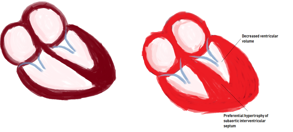
A normal heart vs. a heart that has undergone hypertrophic cardiomyopathy.
How does the patient with HCM present? The decreased cardiac output causes pulmonary venous congestion and hence exertional dyspnoea. The hypertrophied heart causes something of an overcrowding of structures within the ventricles. As the ventricles contract, the anterior leaflet of the mitral valve moves towards the septum and this severe outflow obstruction may allow us to hear a harsh systolic murmur.
HCM may even present as angina- the irregular growth of the myocytes is not matched by an equal ratio of angiogenesis, leading to focal areas of ischemia. Other presentations are atrial fibrillation, mural thrombus formation and, like we saw before, sudden cardiac death.
Now that we have diagnosed the patient as having HCM, what do we do? Plan for a cardiac transplantation? Fortunately, HCM can be treated with β-blocking agents which decrease the heart rate and contractility. Surgical removal of the hypertrophied part of the ventricle using a controlled septal infarction method has also been found to be effective.
As you can see, DCM and HCM are both different and similar and can be confused for the more commonly presenting cardiac pathologies. It’s up to us as clinicians to analyse the patient and history and presentation to come up with a plan to treat the patient.
Author: Shruthi Sivakumar
Sources and citations
Robbins and Coltran Pathologic Basis of Disease, 8th edition.