Toxins - Why size doesn't matter
Infectious diseases, the bread and butter of every medico.
While most of them are trivial, some of them are so powerful that they can cause sepsis and death, or worse, wipe out scores of people.
We all know Vibrio cholera causes cholera, Corynebacterium diphtheria causes diphtheria, Clostridium tetani causes tetanus. Pretty obvious from the name, isn't it?
But, picture this:
Seems very disproportionate, but one such microorganism namely Vibrio cholera proved to be fatal to hundreds of people. 578 people to be exact. In the first ever cholera epidemic recorded by English physician, John Snow, the awesome killing power of these tiny organisms was exposed to the world.
So how do these tiny organisms wield such a devastating power on the mighty human beings?
How does infection by certain bacteria manifest as a certain disease? And if the organism is present in the environment around us, why is it that only few people are affected by the disease? Let us find answers to these questions.
First, let’s refresh some basic microbiology. Many a times, mere presence of bacteria does not cause a disease. We are said to be “infected” if we harbour a pathogenic bacteria which is multiplying inside our body. The infection manifests as a “disease” only at few instances and this ability of an infectious agent to cause a disease is called “virulence”.
Bacteria first adhere to a cell, multiply to form large colonies, invade the tissues to reach the blood stream and release toxins that produce the disease. Though adherence, multiplication, invasion and toxin production are all considered to be important virulence factors, it is the ability to produce toxins which is most dangerous of them all.
Some bacteria release their toxins locally or into the circulation. Such toxins are called Exotoxins. Some bacteria have cell wall which is inherently toxic. Such toxins are called Endotoxins and these bacteria set out on a suicide mission to liberate toxins from their own cell wall after their lysis. While Exotoxins are polypeptide molecules made of amino acids, Endotoxins are lipids. This fundamental difference in the chemical composition of these toxins is of tremendous clinical importance.
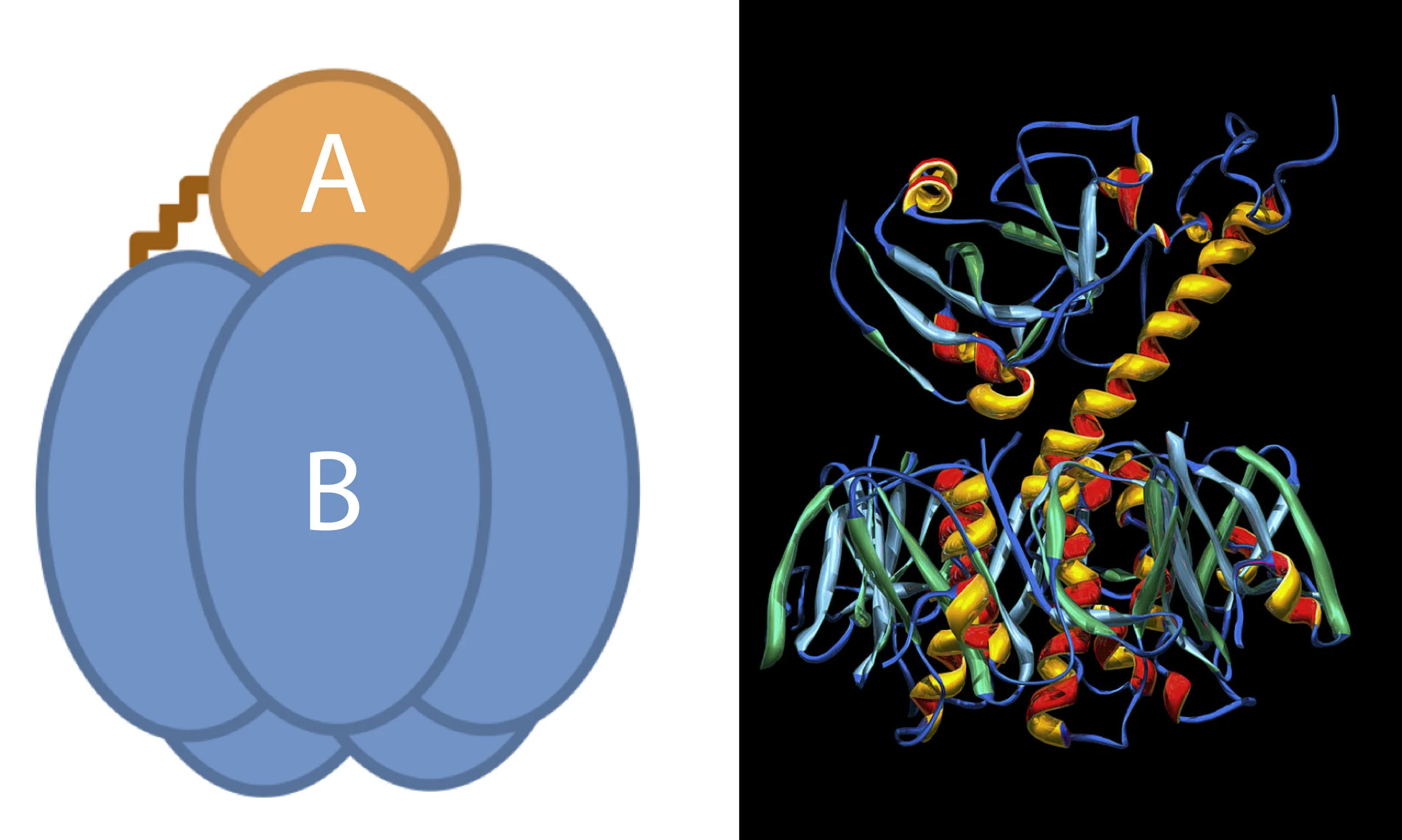
Exotoxins are produced by many Gram positive and Gram negative bacteria. But they all have a similar structure. Exotoxins are made of 2 subunits – A and B. The B subunit helps the toxin to bind to the local tissue and the A subunit attacks the tissue.
Structure of Cholera toxin
Let’s understand this with an example. Cholera is caused by Gram negative bacillus, Vibrio cholera. The bacillus gains access to the intestine after ingesting food contaminated with the bacteria. It then penetrates the intestinal mucosa, multiplies and releases the cholera toxin. B subunit of the toxin firmly binds itself to the mucosal cells. This allows the A subunit to enter the cell and cause an increase in the level of c-AMP which subsequently leads to secretion of massive amounts of electrolytes and fluids into the intestinal lumen causing life threatening diarrhoea.
Since cholera toxin causes diarrhoea, the toxin is also known as Enterotoxin. Examples of other Exotoxins are diphtheria toxin and tetanus toxin.
Did you notice anything common to these 3 diseases?
Yes, diphtheria, tetanus and cholera are some of the vaccine preventable diseases. Since the toxins of these lethal organisms are basically proteins, they undergo denaturation when heated or mixed with formalin. By such methods, Exotoxins can be converted into non virulent products called toxoids and can be used for immunisation. The proteinaceous structure of Exotoxins is also responsible for their extremely lethal nature. A very small quantity of the toxin is sufficient to produce deadly diseases. In fact, tetanus killed more than 50,000 soldiers of the Axis powers in World War II; the Allied forces, however, immunized military personnel against tetanus and very few died of that disease and the rest is history.
In comparison to the Exotoxins, Endotoxins are less toxic. Unlike Exotoxins, Endotoxins are produced only by Gram negative bacteria. You see, the cell wall of Gram negative bacteria is made up of Lipopolysaccharide (LPS) – a complex of lipids and polysaccharide. The lipid component of LPS is known as Lipid A. If you recall from our previous article on lipids, the melting point of a fatty acid increases with increase in the number of carbon atoms (click here for our article on lipids).
Lipid A is decorated with many fatty acids usually having 13 to 18 carbon structures. Due to this unique property of lipids, the LPS molecule as a whole is quite resistant to heat and cannot be converted into non toxic forms, i.e. LPS cannot be toxoided. The death or lysis of Gram negative bacteria like Escherichia coli and Neisseria leads to release of the Endotoxins into the circulation. This causes release of inflammatory mediators like Interleukins leading to varied manifestations ranging from trivial fever to more life threatening complications like disseminated intravascular coagulation, multiple organ failure and death.
And that is how the mighty human being becomes an easy prey to these fascinating, yet sinister creatures!
Author: Soundarya V (Facebook)
Sources and citations
1) Jawetz, Ernest, Edward A. Adelberg, Joseph L. Melnick, and George F. Brooks. "Section 3: Chapter 9: Pathogenesis of Bacterial Infection." Jawetz, Melnick, & Adelberg's Medical Microbiology - - 26. Ed. N.p.: McGraw-Hill Medical, 2013. 155-58. Print.
2) Matsuura, Motohiro. "Structural Modifications of Bacterial Lipopolysaccharide That Facilitate Gram-Negative Bacteria Evasion of Host Innate Immunity." N.p., 2013. Web.N.p.: n.p., n.d. Print.
3) Shiode, Narushige, Shino Shiode, Elodie Rod-Thatcher, Sanjay Rana, and Peter Vinten-Johansen. "The Mortality Rates and the Space-time Patterns of John Snow’s Cholera Epidemic Map." International Journal of Health Geographics. N.p., 2015. Web.


